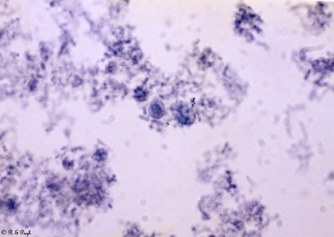
Taxonomy: f. Retortamonadidae
Animal: Retortamonas intestinalis 1 04 .jpg
Sites: Gut
Comment:
Retortamonas intestinalis trophozoites and cyst in faecal smear stained with FeHx. Trophozoite is pear shaped or oval 4 - 9 microns (average 6 - 7) x 3 - 4 microns with 1 nucleus and 1 anterior flagellum and 1 posterior flagellum and a prominent cytostome 1/2 of body; it has a jerky movement in saline wet preparations. Cyst is 4 - 9 x 5 microns pear or lemon shaped with 1 nucleus and outline of cytostome with supporting fibrils. These were found in a 54 year old patient with a thyroid condition and presenting with diarrhoea. She lived in a hippy commune in Australia and been to India 2 years previously. Also present in the faeces were Blastocytis hominis adn C.mesnili but no pathogenic organisms. She was treated with Metronidazole for 7 days, following which symptoms abated although B.hominis (but not R.intestinalis or C.mesnili) were still present in the faeces 7 days and on follow up 4 months later. R.intestinalis is considered non- pathogenic but because of its small size, it is often missed. If the faeces is not examined immediately to see motile trophozoites OR fixed within 1/2 hour of excretion with PVA fixative, the parasites decay. It is essential for identification to examine stained (FeHX or trichrome) smear and preferably 3 specimens collected on alternate days, because if not, there is little chance of detecting these parasites and hence little is known about them. (see Garcia and Bruckner 1997- Bibliography) The pathogenicity of Blastocystis is uncertain. (see Boreham and Stenzel 1993, Stenzel and Boreham 1991 and Stenzel and Boreham 1996 in Bibliography)
First Picture |
Previous Picture |
Next Picture |
Last Picture

